Biomechanics at the Scale of the Cell and Below
Using Laser Surgery
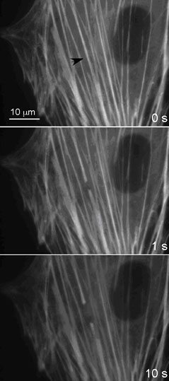
Femtosecond laser illumination can be used to perform surgery at the nano-scale. It can be used to cut structural elements within the cell, without disrupting the surrounding material. The cell at the left has been transfected with a fluorescent label to visualize the actin stress fibers, key structural elements within a cell. When an individual fiber is incised with the laser scalpel, the fiber pulls away from the incision, and contracts. This work from a collaboration between Ingber and Mazur proves that the stress fiber is under tension and permits quantitative analysis of the biomechanical properties of individual stress fibers within the cytoplasm of living cells, something that was never possible in the past. These results are supported by complimentary work by Weitz, showing that actin networks are under tension. Moreover, additional experiments from a collaboration between Weitz and Ingber suggest that the microtubules, a second key stress-bearing structure in cells, are under compression; this will help balance the tension of the actin stress fibers. This provides critical insight into the mechanical properties of the cell.
David A. Weitz (Physics & Applied Physics)
D. Ingber, and
E. Mazur
Harvard MRSEC (DMR-0820484)